I build organoids, murine and human, and the tools to read them.
Self-organizing tissue models combined with quantitative image analysis, providing experimental systems that generate reproducible drug-response data at the preclinical stage.
- Murine and human organoid models that recapitulate tissue physiology in a dish
- High-throughput image analysis that turns high-resolution microscopy into quantitative data
- New Approach Methodologies (NAMs) that reduce, refine and where possible replace animal use
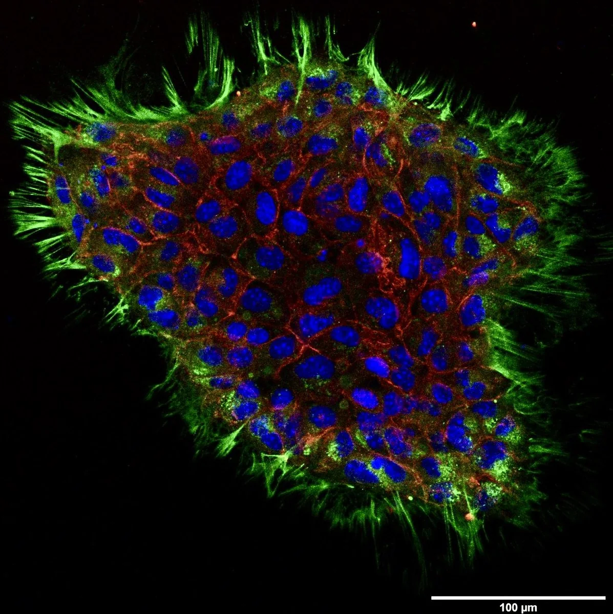
Dissecting disease and testing therapeutics in in vitro models.
In April 2025 the U.S. FDA issued its Roadmap to Reducing Animal Testing in Preclinical Safety Studies, formally positioning New Approach Methodologies (NAMs, including organ-on-chip systems, in silico toxicology, and human cell-based assays) as a route to reduce, refine and potentially replace animal testing in preclinical safety studies, beginning with monoclonal antibodies1. The context is attrition: of the candidates that enter phase I, only about one in ten is eventually approved3, and the largest single cause of late-stage failure is insufficient efficacy, followed by safety, with a further share of programmes discontinued on strategic or commercial grounds4,5. The agency's own stated rationale is that animal studies are a limited predictor of how a drug performs in humans2; animal models therefore contribute to that gap wherever human-specific pharmacology and safety liabilities cannot be extrapolated across species, rather than accounting for it in full.
My research is aligned with this trend. I build organoid models that preserve native tissue architecture (intestinal and colonic epithelial organoids derived from mouse adult stem cells, and human iPSC-derived vascular organoids), coupled with high-throughput image analysis that turns multi-layered images into reproducible, operator-independent readouts. The same platform can be applied across multiple contexts: as a mechanistic disease model, as a high-throughput system for compound screening and safety assessment, and, with patient-derived specimens, as an experimental basis for personalised therapeutic evaluation.
The transcriptional logic of collective invasion
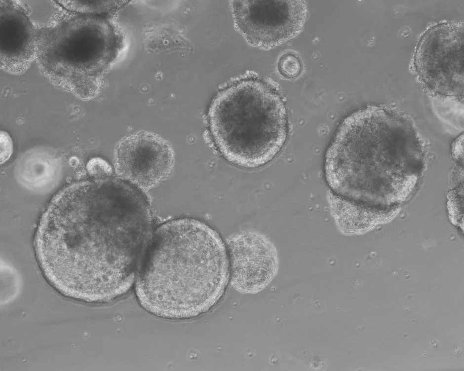
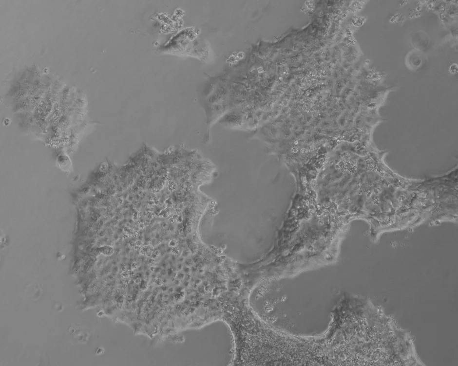
Colorectal carcinoma cells frequently invade not as single migratory cells but as cohesive collectives undergoing a partial epithelial–mesenchymal transition (pEMT), retaining cell–cell contacts while acquiring mesenchymal motility. Using oncogenically transformed murine intestinal organoids (derived from adult intestinal stem cells) as a tractable model of carcinogenesis, my doctoral work identified the transcription factor Sox11 as necessary, though not sufficient, for this program.
Canonical TGF-β1 signaling redirects Sox11's gene-regulatory activity: Sox11-dependent genes are enriched for EMT, TGF-β and PDGF pathway components, and their human orthologues co-vary with SOX11 in colorectal cancer.
The study combined single-cell and bulk RNA-sequencing, gene-regulatory-network inference (SCENIC), and more than twenty CRISPR-Cas9 loss- and gain-of-function lines for functional validation. It established a mechanistic framework (published as first author in Oncogenesis, 2025) describing how a single transcription factor is repurposed by the tumour microenvironment to promote invasion.
Perturbing and visualizing tissue in situ
Mechanistic claims require perturbation. For loss-of-function, I generate CRISPR-Cas9 knockouts by dual-sgRNA exon deletion, then isolate single-cell-derived clonal lines validated by PCR and Sanger sequencing; across the project this amounts to more than twenty knockout and overexpression lines, each coupled to functional readouts. Beyond loss- and gain-of-function, I apply CRISPR-HOT (homology-independent organoid transgenesis) to knock fluorescent reporters, epitope tags, and degron cassettes (including a mutant FKBP degron for conditional protein destabilisation) in frame at endogenous loci.
This makes it possible to visualize and acutely control transcription factors at the invasive front of living organoids, linking molecular intervention to morphological consequence in the same intact tissue.
Blood vessel organoids for drug screening
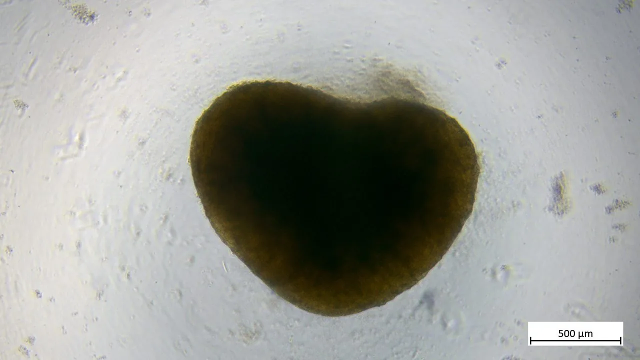
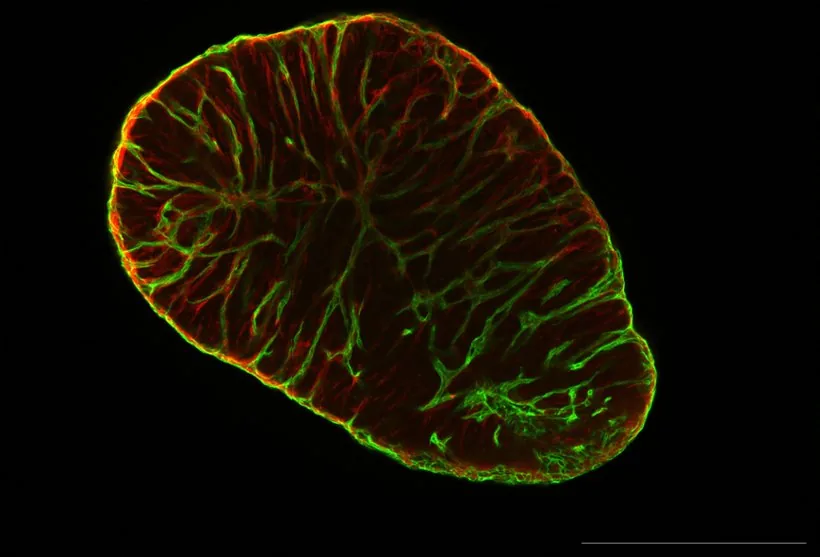
Human iPSC-derived blood vessel organoids self-assemble interconnected networks of endothelial cells and pericytes enclosed by a basement membrane, forming three-dimensional human vascular tissue; the platform builds on the protocol of Wimmer et al. (Nature 2019;565:505–510). At Angios FlexCo, this work established high-throughput production pipelines across multiple hydrogel embedding conditions, scaling these organoids from research-scale preparation toward a screening-ready platform.
On top of production, the work established a quantitative vascular toxicity workflow comprising compound treatment, immunofluorescence staining, confocal acquisition, and automated readout of vascular morphology and signal intensity. The pipeline supports dose-response characterization of vascular-disrupting and anti-angiogenic agents, consistent with the regulatory and ethical basis for New Approach Methodologies (NAMs) that reduce reliance on animal models.
A methods contribution describing an animal-origin-free route to these organoids is co-authored and published in Scientific Reports (2026).
Quantitative readouts from confocal image stacks
The utility of a model depends on the quality of the readouts it produces. This work develops automated image-analysis pipelines in Python and ImageJ macros to reduce subjectivity in organoid phenotyping. For vascular organoids, this includes segmentation, skeletonization, and graph-based network analysis (via networkx) to quantify branch topology, together with morphometric descriptors such as an approximate average diameter, taken as the mean of the maximum and minimum Feret diameters rather than the true angular mean.
For marker localization, a dedicated macro quantifies radial intensity profiles of endothelial (CD31) and pericyte (PDGFRB) signals, capturing spatial organization that simple intensity averages would miss. Every pipeline is written to fixed, documented conventions so that results are reproducible across experimental runs and transferable to collaborators.
How the work gets done.
The experimental and computational toolkit behind my doctoral work: build a faithful disease model, perturb it with genome engineering, read it out by imaging and sequencing, then anchor every finding back to patients.
TKA Organoid Model
A mouse intestinal organoid carrying the three most common colorectal-cancer driver mutations (Apc, KrasG12D, Trp53R172H), switchable on demand.
CRISPR Knockout · Clonal Lines
Deleting a gene cleanly from an organoid and growing single-cell-derived clones to ask what that gene was for.
CRISPR-HOT Endogenous Tagging
Marking a protein at its own gene so it can be seen and degraded, when no usable antibody exists.
Inducible Overexpression
Switching a gene on with a drug to test whether it is sufficient, the gain-of-function counterpart to knockout.
Transwell Invasion Assay
Turning collective invasion into a number, so that genotypes can be compared quantitatively.
Whole-Mount Immunofluorescence
Imaging proteins in an intact 3D organoid by confocal microscopy, to see not just how much but where.
Single-Cell RNA-seq + Trajectory
Reading every cell in a treated organoid separately, then ordering them along the path from epithelial to invasive.
Regulon & Leader-Cell Analysis
Inferring which transcription factor controls the invasive cells, and scoring which cells are leaders.
Bulk RNA-seq
Deep population-level transcriptomes to validate the single-cell findings and define gene modules.
CRC Patient Data Mining
Testing whether an organoid finding holds in real patients, using public cancer-genomics and survival data.
iPSC Vascular Organoid · HTP Generation
Differentiating human iPSCs into self-organizing blood-vessel organoids (endothelium and pericytes), and scaling the protocol into high-throughput plate formats for screening.
Drug Testing · IF, Confocal & Network Analysis
Treating vascular organoids with compounds, then reading structural damage by immunofluorescence, confocal imaging, and quantitative vascular-network analysis (CD31 / PDGFRB; length, branching, diameter) to score vascular toxicity.
A researcher integrating experimental biology and computation.
Since 2015, work across academia and industry developing advanced in vitro models, ranging from the molecular mechanisms of cancer to screening-ready human tissue platforms.
I am a PhD scientist working on organoids (three-dimensional tissue models spanning murine adult-stem-cell-derived intestinal and colonic epithelium and human iPSC-derived blood vessels) and the computational analytics required to turn them into reliable experimental systems. My trajectory has moved from fundamental discovery toward translational application, since the most useful organoid platforms are generally built where rigorous biology meets reproducible quantification.
My doctoral research at the University of Freiburg, in the Andreas Hecht lab, examined how colorectal cancer cells invade collectively. I identified the transcription factor Sox11 as a necessary, though not sufficient, regulator of partial EMT, integrating single-cell transcriptomics, gene-regulatory-network inference, and extensive CRISPR perturbation. This work established an approach of pairing mechanistic hypotheses with the analytical infrastructure required to test them at scale.
A model that cannot be quantified reproducibly is insufficient as a reliable experimental system.
At Angios FlexCo in Innsbruck, I developed high-throughput vascular organoid platforms and drug-toxicity workflows; this direction is consistent with the field's move toward New Approach Methodologies (NAMs).
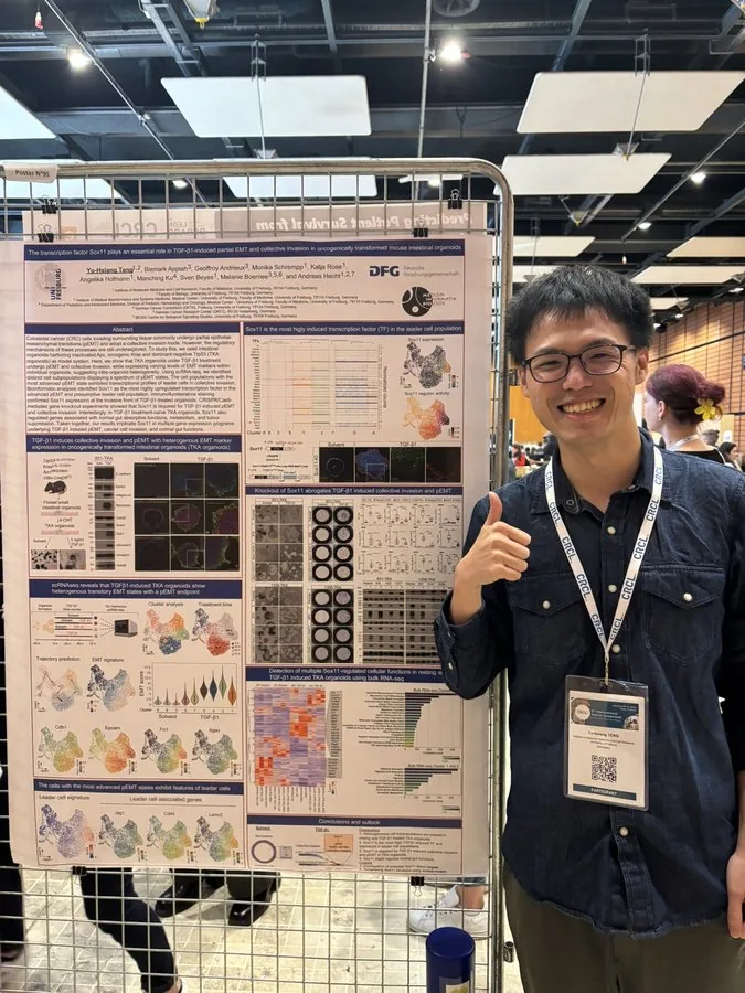
Outside the lab I am a constant traveler (more than twenty countries across Asia and Europe), a competitive badminton player of eight years, and an alpine hiker who logs over twenty trails a year in the Alps and the Black Forest. In Europe, I cook seriously in the Chinese and Japanese traditions. These habits reflect my attention to detail, endurance, and adaptability.
Let's build a better model.
Open to collaborations in organoid platform development, preclinical screening, NAMs, and quantitative image analysis across academic, CRO, and pharmaceutical settings.
Relocating from Innsbruck to Taiwan in August 2026 and joining a preclinical CRO in New Taipei City as Senior Scientist from September; recent work centered on high-throughput vascular organoid and drug-toxicity platforms.
Platform Development
Standing up iPSC-derived organoid systems and high-throughput culture workflows from protocol to reproducible pipeline.
Screening & NAMs
Designing drug-response and toxicity assays on human organoid models aligned with New Approach Methodologies.
Image Analytics
Automated quantification of organoid morphometry, marker intensity, and vascular network topology in Python & ImageJ.
Reading the field, and placing my own work in it.
A year-by-year read of the organoid literature from 2021 to 2026, and the platform I am building from it.
Five years of the field
An organoid is a self-organising 3D mini-tissue grown from pluripotent (iPSC/ESC) or adult stem cells, sitting between architecture-poor 2D cell lines and species-divergent animal models. Reading the 2021 to 2026 literature, the same three directions keep coming up: closing the gap to in-vivo maturity, engineering in more complexity (vascularisation, immune compartments, assembloids), and building the functions that make a model useful for translation.
Stem-cell embryo models and patterned tissue
Stem cells self-organise into increasingly faithful early-development and tissue-specific structures. Human blastoids reconstitute blastocyst-stage architecture for the ethical study of implantation and lineage segregation, while directed differentiation yields patterned kidney organoids that recapitulate the adult collecting-duct system.
- Human blastoids model blastocyst development and implantation — Kagawa et al. (Rivron lab); Nature 2022;601:600–605 (online 2021).
- Generation of patterned kidney organoids that recapitulate the adult kidney collecting duct system from expandable ureteric bud progenitors — Zeng et al.; Nat Commun 2021;12:3641.
- SARS-CoV-2 productively infects human gut enterocytes — Lamers et al. (Clevers lab); Science 2020;369:50–54.
In-vivo maturation and matrix alternatives
Transplantation into a living host solved the maturation ceiling: human cortical organoids grafted into newborn rat cortex vascularise and enlarge about ninefold in volume over three months post-transplantation, integrate into sensory and motivation circuits, and expose patient-specific (e.g. Timothy-syndrome) deficits invisible in a dish. In parallel, tissue-derived and synthetic ECM hydrogels emerged as alternatives to animal-derived Matrigel.
- Maturation and circuit integration of transplanted human cortical organoids — Revah et al. (Pașca lab); Nature 2022;610:319–326.
- A scalable organoid model of human autosomal dominant polycystic kidney disease for disease mechanism and drug discovery — Tran et al.; Cell Stem Cell 2022;29:1083–1101.
- Tissue extracellular matrix hydrogels as alternatives to Matrigel for culturing gastrointestinal organoids — Kim et al.; Nat Commun 2022;13:1692.
Biocomputing, immune integration and high-throughput screening
Brainoware framed living neural tissue as a reservoir-computing substrate, performing speaker identification on a Japanese-vowel dataset (reported by the authors as speech recognition) and non-linear prediction through a high-density multielectrode array. iPSC-derived microglia added a functional innate-immune compartment that drives maturation, and bioprinting with label-free interferometry pushed drug screening to single-organoid resolution.
- Brain organoid reservoir computing for artificial intelligence — Cai et al. (Guo lab); Nat Electron 2023;6:1032–1039.
- iPS-cell-derived microglia promote brain organoid maturation via cholesterol transfer — Park et al.; Nature 2023;623:397–405.
- Drug screening at single-organoid resolution via bioprinting and interferometry — Tebon et al.; Nat Commun 2023;14:3168.
Cell therapy and human-relevant safety pharmacology
hPSC-derived intestinal organoids engrafted and repaired damaged bowel in a rodent injury model, a regenerative-medicine rather than a NAMs milestone. For safety, donor-matched intestinal organoids and tumouroids quantified the on-target / off-tumour toxicity of T-cell-engaging bispecific antibodies — the human-relevant question NAMs aim to answer without animals; decellularised-tissue (natural) hydrogels supported vascularised kidney organoids.
- Analysis of off-tumour toxicities of T-cell-engaging bispecific antibodies via donor-matched intestinal organoids and tumouroids — Harter et al.; Nat Biomed Eng 2024;8:345–360.
- Human pluripotent stem cell-derived organoids repair damaged bowel in vivo — Poling et al.; Cell Stem Cell 2024;31:1513–1523.e7.
- Natural hydrogels support kidney organoid generation and promote in vitro angiogenesis — Garreta et al.; Adv Mater 2024;36(34):e2400306.
Metabolic, vascular and immune function
Metabolically competent human adult hepatocyte organoids (Wnt/STAT3-driven self-renewal; CYP450 activity, albumin/urea and bile handling) supply a prerequisite for animal-free drug-induced liver-injury (DILI) assessment. Vascularised gastruloids modelled the earliest human cardiac and hepatic vascularisation, and blood-derived synthetic immune organoids reconstituted functional germinal-centre B-cell responses.
- Generation of human adult hepatocyte organoids with metabolic functions — Igarashi et al. (Sato lab); Nature 2025;641:1248–1257.
- Gastruloids enable modeling of the earliest stages of human cardiac and hepatic vascularization — Abilez et al. (Wu lab); Science 2025;388:eadu9375.
- Human immune organoids to decode B cell response in healthy donors and patients with lymphoma — Zhong et al. (Singh lab); Nat Mater 2025;24:297–311.
AI-augmented and automated organoids
The field is increasingly combining AI and automation. AI virtual organoids (organoid-scale digital twins) propose in-silico counterparts that reduce wet-lab runs; method reviews formalise machine-learning and mechanistic-modelling pipelines; and robotic, miniaturised PDO screening addresses the throughput and reproducibility gaps that have limited adoption.
- Artificial Intelligence Virtual Organoids (AIVOs) — Bai & Su; Bioact Mater 2026;59:45–68 (online 2025).
- ML, AI & mathematical modelling for organoid research — Trends in Biotechnology 2026.
- Automated, miniaturised PDO drug screening — npj Biomedical Innovations 2026.
Representative studies are illustrative, not an exhaustive or quantitative ranking. Titles are quoted verbatim and citations follow the volume year of record, with the online-first year given where the two differ; entries for 2021 to 2025 were checked against the primary sources, and two 2026 entries are cited at journal level only.
My roadmap
These trends are the direction I'm taking. My earlier work on intestinal organoids and CRISPR was a mechanistic disease model; more recently I moved to building iPSC organoid platforms and their toxicity readouts, the human-relevant safety testing the 2024 and 2025 studies point to; next is the reproducibility and throughput the 2026 automation work aims to solve. NAMs is converging on models that are human-relevant, quantifiable and screening-ready, and that is the platform I am building toward.